Position of the Heart in the Thoracic Region
Colour by Numbers Activity
The content in this activity aligns with Chapter 9: Cardiovascular System – Heart in Building a Medical Terminology Foundation 2e.
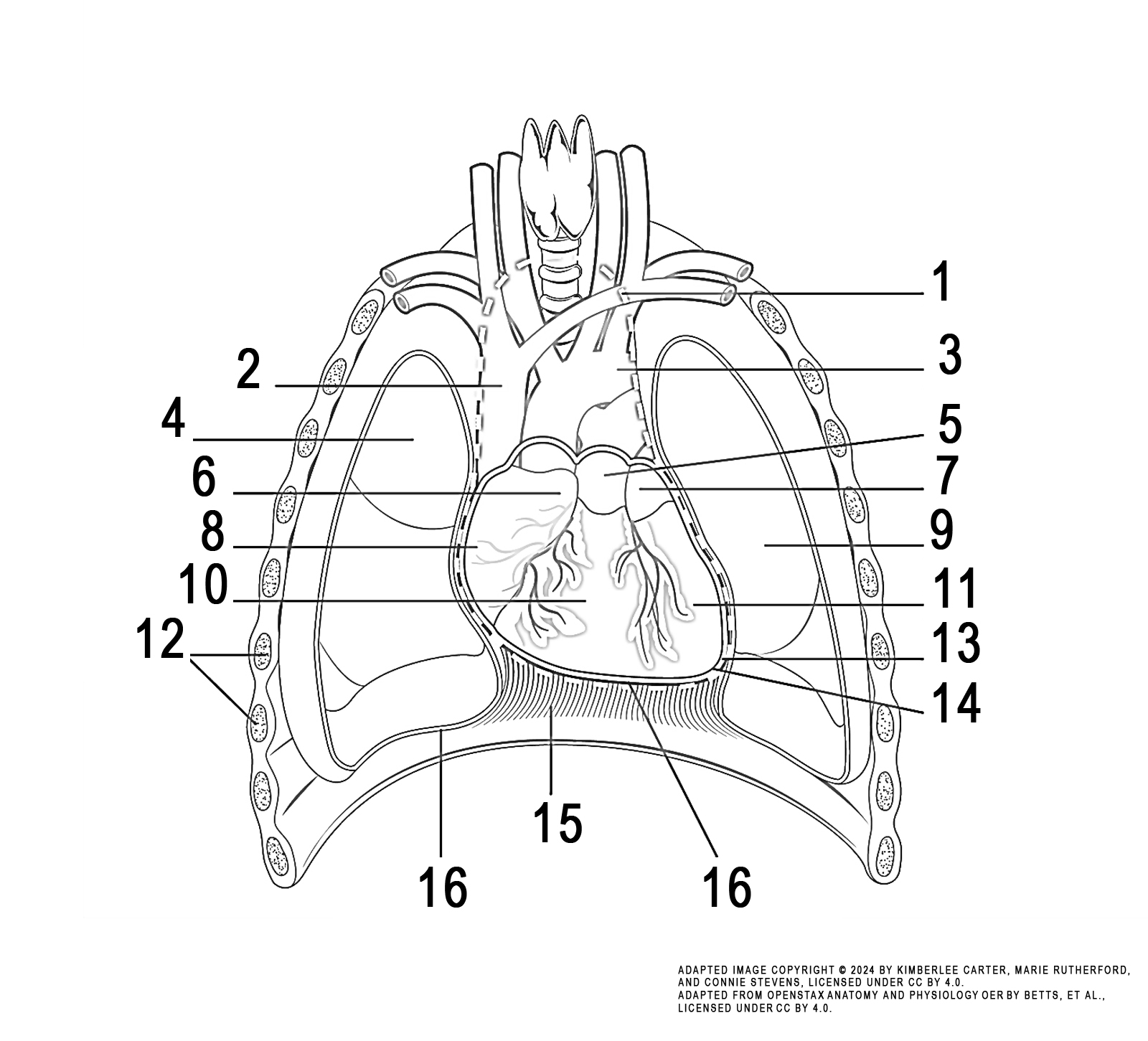
Instructions: Review the illustration. This illustration displays an anterior view of the anatomy and positioning of the heart.
Task: Colour each numbered structure using the colour indicated in the list below:
- mediastinum (orange)
- superior vena cava (dark blue)
- arch of aorta (red)
- right lung (green)
- pulmonary trunk (light blue)
- right auricle (grey)
- left auricle (yellow)
- right atrium (purple)
- left lung (red)
- right ventricle (pink)
- left ventricle (brown)
- ribs (yellow)
- pericardial cavity (grey)
- apex of the heart (pink)
- diaphragm (green)
- edge of the parietal pleura/pericardium – both sides (purple)

Image Attribution
The image used in this activity was sourced from OpenStax Anatomy and Physiology OER by Betts, et al., which is licensed under CC BY 4.0. Following OpenStax’s leadership and in the spirit of open education, we have licensed this OER with the same license.
Image Description
This illustration activity shows the location of the heart in the thorax. The anterior view includes (from top, clockwise): mediastinum, arch of aorta, pulmonary trunk, left auricle, left lung, left ventricle, pericardial cavity, apex of heart, edge of parietal pericardium, diaphragm, edge of parietal pleura, ribs, right ventricle, right atrium, right auricle, right lung, superior vena cava.
