Posterior Pituitary
Colour by Numbers Activity
The content in this activity aligns with Chapter 17: Endocrine System in Building a Medical Terminology Foundation 2e.
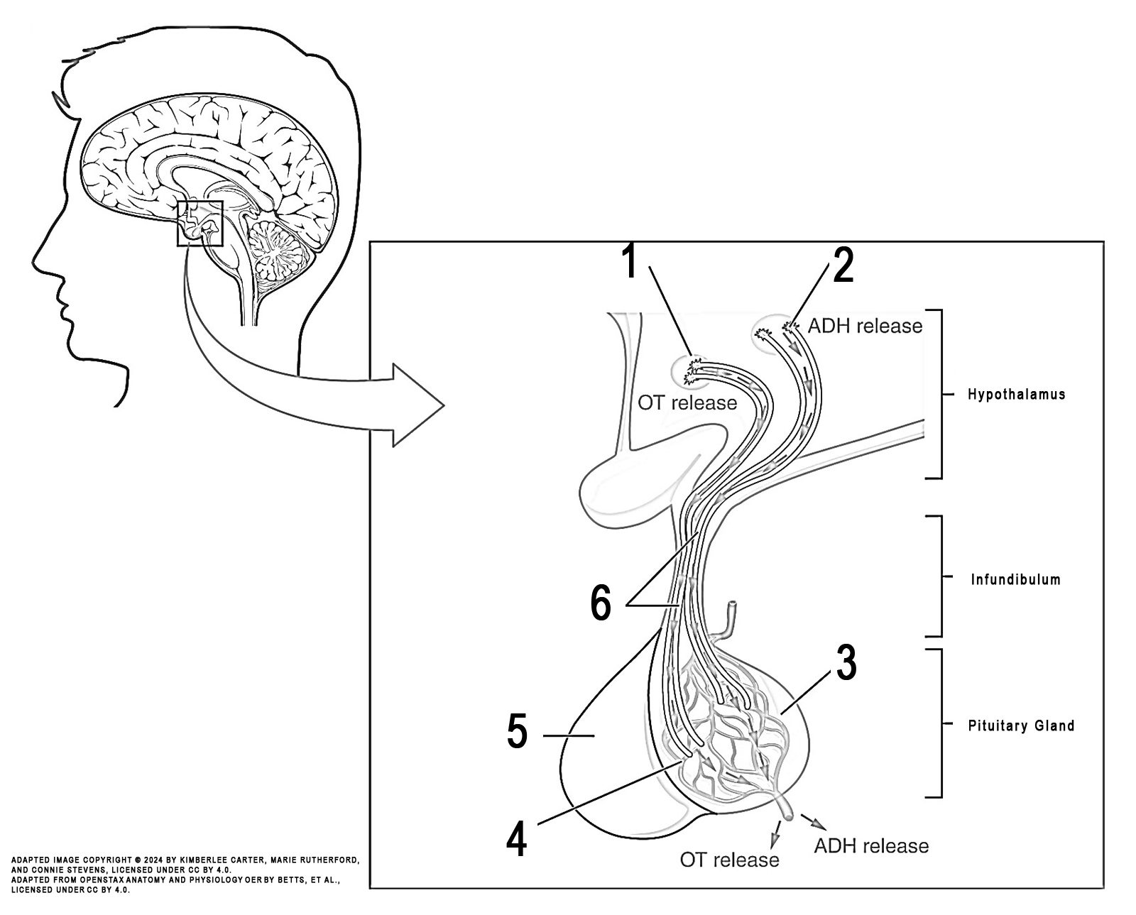
Instructions: Review the illustration. This illustration displays the posterior pituitary gland.
Task: Colour each numbered structure using the colour indicated in the list below:
- neurosecretory cell of supraoptic nucleus (green)
- neurosecretory cell of supraoptic nucleus (orange)
- posterior pituitary (yellow)
- capillary plexus (grey)
- anterior pituitary (purple)
- hypothalamohypophyseal tract (green)

Image Attribution
The image used in this activity was sourced from OpenStax Anatomy and Physiology OER by Betts, et al., which is licensed under CC BY 4.0. Following OpenStax’s leadership and in the spirit of open education, we have licensed this OER with the same license.
Image Description
This illustration activity shows the posterior pituitary. This illustration zooms in on the hypothalamus and the attached pituitary gland. The posterior pituitary is highlighted. Two nuclei in the hypothalamus contain neurosecretory cells that release different hormones. The neurosecretory cells of the paraventricular nucleus release oxytocin (OT), while the neurosecretory cells of the supraoptic nucleus release anti-diuretic hormone (ADH). The neurosecretory cells stretch down the infundibulum into the posterior pituitary. The tube-like extensions of the neurosecretory cells within the infundibulum are the hypothalamophypophyseal tracts. These tracts connect with a web-like network of blood vessels in the posterior pituitary called the capillary plexus. From the capillary plexus, the posterior pituitary secretes the OT or ADH into a single vein that exits the pituitary.
