Accessory Structure of Skin – Hair
Colour by Numbers Activity
The content in this activity aligns with Chapter 3: Integumentary System in Building a Medical Terminology Foundation 2e.
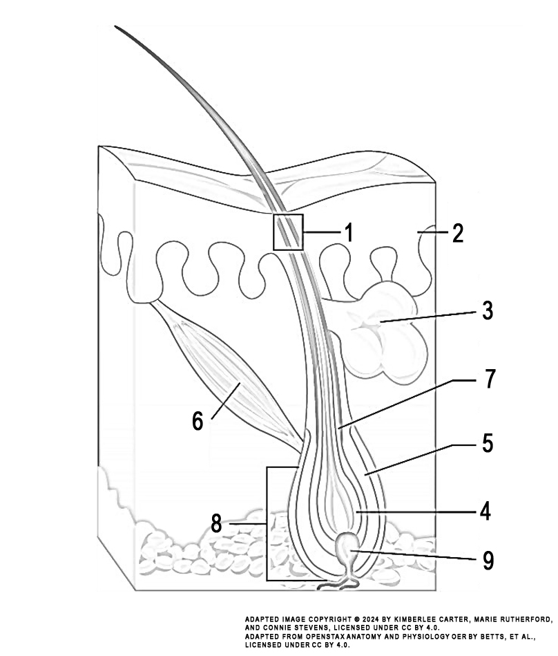
Instructions: Review the illustration. This illustration displays the structures making up a hair follicle.
Task: Colour each numbered structure using the colour indicated in the list below:
- hair shaft (green)
- medulla, cortex, cuticle (red)
- sebaceous gland (dark blue)
- hair matrix (brown)
- outer root sheath (orange)
- arrector pili muscle (purple)
- inner root sheath (pink)
- hair bulb (grey)
- hair papilla (light blue)

Image Attribution
The image used in this activity was sourced from OpenStax Anatomy and Physiology OER by Betts, et al., which is licensed under CC BY 4.0. Following OpenStax’s leadership and in the spirit of open education, we have licensed this OER with the same license.
Image Description
This illustration activity shows a cross section of the skin containing a hair follicle. The follicle is teardrop shaped. Its enlarged base, labeled the hair bulb, is embedded in the hypodermis. The outermost layer of the follicle is the epidermis, which invaginates from the skin surface to envelope the follicle. Within the epidermis is the outer root sheath, which is only present on the hair bulb. It does not extend up the shaft of the hair. Within the outer root sheath is the inner root sheath. The inner root sheath extends about half of the way up the hair shaft, ending midway through the dermis. The hair matrix is the innermost layer. The hair matrix surrounds the bottom of the hair shaft where it is embedded within the hair bulb. The hair shaft, in itself, contains three layers: the outermost cuticle, a middle layer called the cortex, and an innermost layer called the medulla.
