Nail Structure
Colour by Numbers Activity
The content in this activity aligns with Chapter 3: Integumentary System in Building a Medical Terminology Foundation 2e.
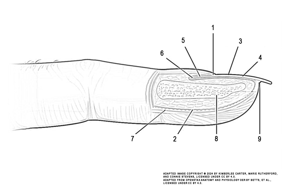
Instructions: Review the illustration. This illustration displays the structures making up the nail.
Task: Colour each numbered structure using the colour indicated in the list below:
- eponychium (green)
- dermis (red)
- lunula (dark blue)
- nail body (brown)
- proximal nail fold (orange)
- nail root (purple)
- epidermis (pink)
- phalanx (grey)
- hyponychium (light blue)

Image Attribution
The image used in this activity was sourced from OpenStax Anatomy and Physiology OER by Betts, et al., which is licensed under CC BY 4.0. Following OpenStax’s leadership and in the spirit of open education, we have licensed this OER with the same license.
Image Description
This illustration activity shows the anatomy of the fingernail region. The image shows a lateral view of the nail bed anatomy. The proximal nail fold is the part underneath where the skin of the finger connects with the edge of the nail. The eponychium is a thin, pink layer between the white proximal edge of the nail (the lunula), and the edge of the finger skin. The lunula appears as a crescent-shaped white area at the proximal edge of the pink-shaded nail. The lateral nail folds are where the sides of the nail contact the finger skin. The nail grows distally out from the proximal nail fold. The edge of the nail is located just proximal to the nail fold. This end of the nail, from which the nail grows, is called the nail root.
