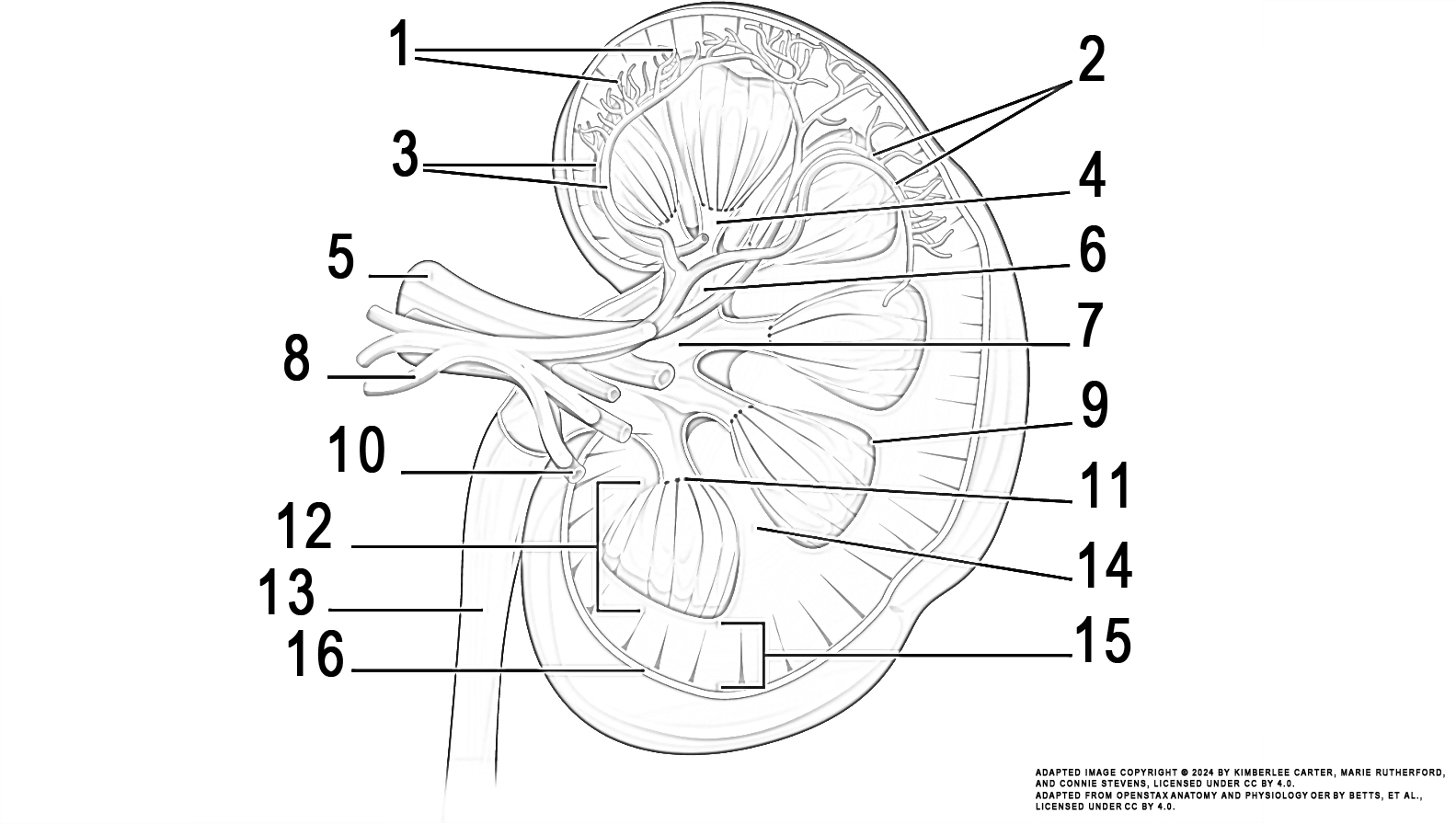
Left Kidney
Colour by Numbers Activity
The content in this activity aligns with Chapter 5: Urinary System in Building a Medical Terminology Foundation 2e.
Instructions: Review the illustration. This illustration displays the anatomy and structures comprising the kidney.
Task: Colour each numbered kidney structure using the colour indicated in the list below:
- cortical blood vessels (orange)
- arcuate blood vessels (red)
- interlobular blood vessels (brown)
- minor calyx (green)
- renal vein (dark blue)
- major calyx (grey)
- renal pelvis (yellow)
- renal hilum and renal nerve (light blue)
- pyramid (purple)
- renal artery (red)
- papilla (pink)
- medulla (brown)
- ureter (yellow)
- renal column (grey)
- outer root sheath (orange)
- cortex (green)
- capsule (purple)

Image Attribution
The image used in this activity was sourced from OpenStax Anatomy and Physiology OER by Betts, et al., which is licensed under CC BY 4.0. Following OpenStax’s leadership and in the spirit of open education, we have licensed this OER with the same license.
Image Description
This illustration activity shows a cross section of the left kidney. There is an outer region called the renal cortex and an inner region called the medulla. The renal columns are connective tissue extensions that radiate downward from the cortex through the medulla to separate the most characteristic features of the medulla, the renal pyramids and renal papillae. The papillae are bundles of collecting ducts that transport urine made by nephrons to the calyces of the kidney for excretion. The renal columns also serve to divide the kidney into 6–8 lobes and provide a supportive framework for vessels that enter and exit the cortex. The pyramids and renal columns taken together constitute the kidney lobes.
