Pericardial Membranes and Layers of the Heart
Highlight/Shading Activity
The content in this activity aligns with Chapter 9: Cardiovascular System – Heart in Building a Medical Terminology Foundation 2e.
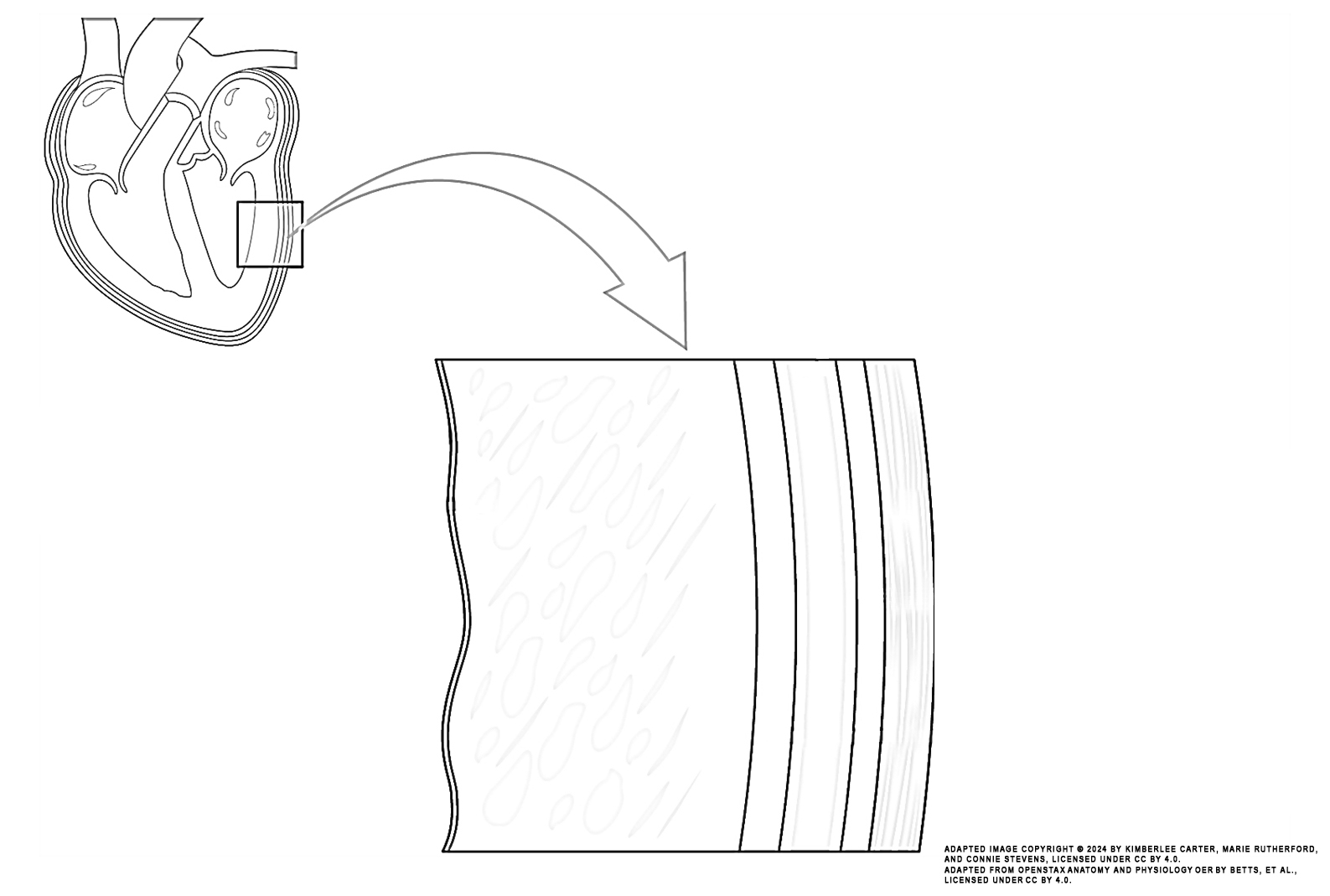
Instructions: Review the illustration. The structural layers of the heart are identified.
Task: Outline the layers of the heart by shading it or highlighting them in the colour shown in the list:
- pericardial cavity (dark blue)
- fibrous pericardium (green)
- parietal layers of serous pericardium (pink)
- epicardium visceral layer (yellow)
- endocardium and myocardium (red)

Image Attribution
The image used in this activity was sourced from OpenStax Anatomy and Physiology OER by Betts, et al., which is licensed under CC BY 4.0. Following OpenStax’s leadership and in the spirit of open education, we have licensed this OER with the same license.
Image Description
This illustration activity shows a magnified view of the structure of the heart wall. Structures include (from top, clockwise): pericardial cavity, fibrous pericardium, parietal layer of serous pericardium, epicardium (visceral layer of serous pericardium), myocardium, endocardium.
